
Echocardiography (Echo)
Echocardiography (commonly called Echo) is a non-invasive ultrasound-based imaging technique used to visualize the heart's structure and function in real time. It evaluates chamber size, valve function, wall motion, and blood flow.
What We Evaluate:
- Chamber Size - Heart chamber dimensions
- Valve Function - All cardiac valves
- Wall Motion - Contractility assessment
- Blood Flow - Flow patterns and velocity
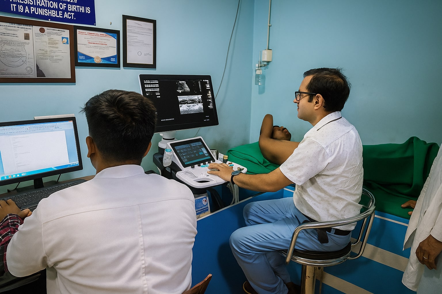
How Echocardiography Works
Ultrasound-based real-time cardiac imaging
High-Frequency Sound Waves
Uses high-frequency sound waves (ultrasound) directed at the heart
Sound Wave Reflection
The probe (transducer) sends sound waves that bounce off heart structures
Real-Time Images
Returning echoes are converted into images or videos on a screen
What Echo Evaluates
Comprehensive cardiac assessment
Chamber Size
Assessment of heart chamber dimensions
- Left ventricle size
- Right ventricle size
- Atrial dimensions
- Chamber volumes
Valve Function
Comprehensive valve assessment
- Mitral valve
- Aortic valve
- Tricuspid valve
- Pulmonary valve
Wall Motion
Cardiac wall movement evaluation
- Regional wall motion
- Contractility assessment
- Ischemia detection
- Scar tissue identification
Blood Flow
Blood flow patterns and velocity
- Doppler flow assessment
- Regurgitation detection
- Stenosis evaluation
- Cardiac output
Types of Echocardiography
Multiple imaging modalities for comprehensive assessment
2D Echocardiography
Two-dimensional images of heart structures
- Standard heart imaging
- Valve visualization
- Chamber assessment
- Basic function evaluation
Doppler Echocardiography
Measures blood flow speed and direction
- Blood flow velocity
- Valve stenosis/regurgitation
- Pressure gradients
- Cardiac output measurement
Color Flow Imaging
Shows blood flow in color for better visualization
- Visual flow patterns
- Leak detection
- Regurgitation assessment
- Flow direction
3D Echocardiography
Three-dimensional heart structure visualization
- Detailed valve anatomy
- Volume assessment
- Complex defect evaluation
- Surgical planning
Advantages of Echocardiography
Safe, effective, and comprehensive cardiac imaging
Non-Invasive
Completely painless ultrasound-based procedure
Real-Time Imaging
Live visualization of heart structure and function
No Radiation
Safe ultrasound technology with no radiation exposure
Comprehensive Assessment
Evaluates chambers, valves, walls, and blood flow
Quick Procedure
Typically takes 30-60 minutes to complete
Repeatable
Can be done multiple times for monitoring
Cardiac Conditions Detected
Comprehensive diagnosis of heart conditions
Your Echo Test Journey
Simple and comfortable cardiac imaging
Preparation
Patient positioned and gel applied
Probe Placement
Transducer placed on chest
Image Acquisition
Multiple views captured
Analysis
Cardiologist reviews and reports
Frequently Asked Questions
Book Your Echocardiogram Today
Non-invasive, real-time cardiac imaging for comprehensive heart assessment