
USG with Colour Doppler
A real-time imaging technique which uses high-frequency sound waves to detect the cause of illness and problems, and find out the actual disease by creating images of organs and internal structures.
What We Image:
- Pregnancy - Safe fetal monitoring
- Abdominal - Organ evaluation
- Cardiological - Heart assessment
- Musculoskeletal - Joint & soft tissue
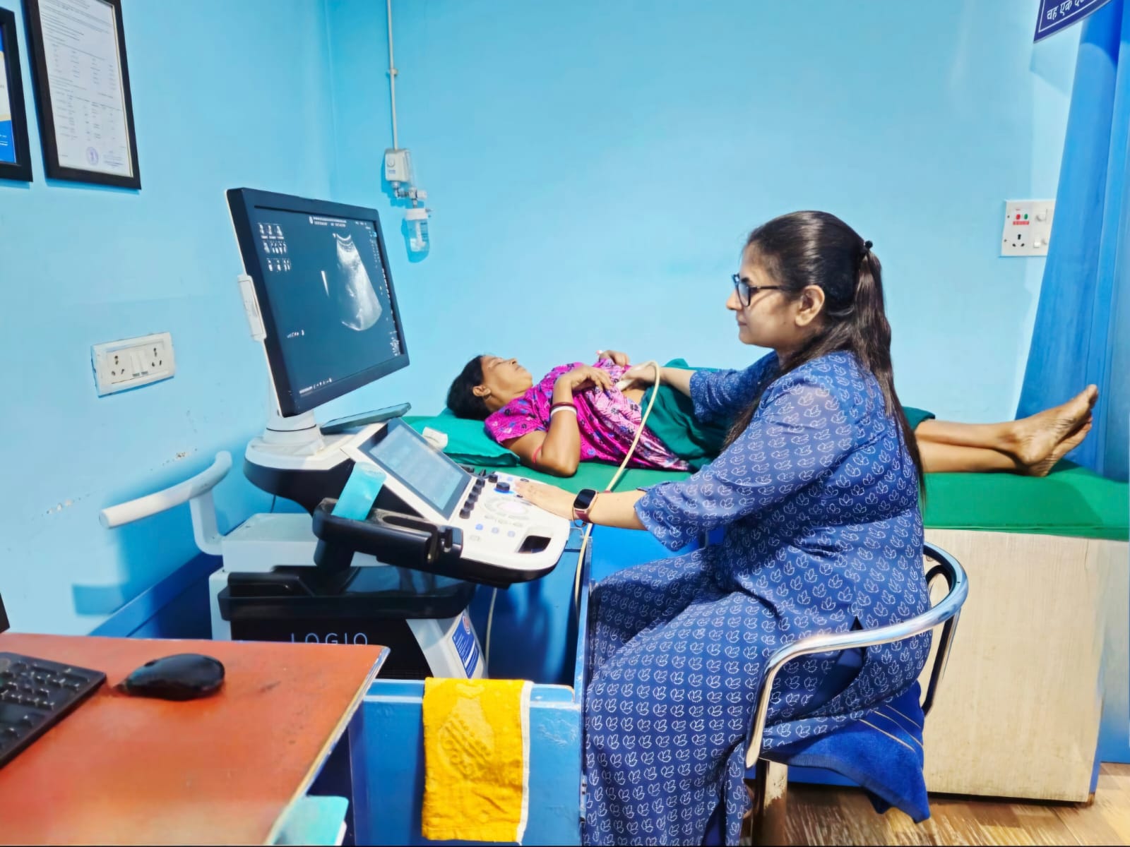
✓ Performed by Specialist Doctors Only
How Ultrasound Works
Safe, non-invasive imaging using sound waves
High-Frequency Sound Waves
Ultrasound probe emits high-frequency sound waves into the body
Echo Detection
Sound waves bounce back from organs and blood flow, creating echoes
Real-Time Imaging
Computer processes echoes to create images showing structure and blood flow
The Procedure
Generally, ultrasound is non-invasive. A special gel is applied to the skin, and a transducer (probe) is moved over the area of interest and need to capture the images of organs. The gel helps eliminate air between the transducer and skin for better image quality.
The entire process is painless and typically takes 15-45 minutes depending on the area being examined.
Types of Ultrasound Imaging
Comprehensive imaging for various medical needs
Pregnancy Imaging
Safe monitoring of fetal development and umbilical cord blood flow
- Fetal growth assessment
- Placental blood flow
- Umbilical cord evaluation
- Pregnancy monitoring
Abdominal Imaging
Comprehensive evaluation of abdominal organs and vessels
- Liver assessment
- Kidney evaluation
- Spleen imaging
- Pancreas examination
Cardiological Imaging
Heart structure and blood flow assessment
- Heart valve function
- Blood flow patterns
- Cardiac chamber evaluation
- Vessel assessment
Musculoskeletal Imaging
Evaluation of muscles, tendons, and soft tissues
- Joint inflammation
- Tendon injuries
- Muscle tears
- Soft tissue evaluation
Advantages of USG with Colour Doppler
Safe, effective, and advanced imaging technology
No Radiation Exposure
Completely safe with no harmful radiation
Real-Time Imaging
Live visualization of organs and blood flow
Non-Invasive Procedure
Simple gel application with transducer movement
Safe for Pregnancy
Totally safe for pregnancy monitoring
Specialist Performed
Conducted by specialist doctors only
Color Doppler Technology
Visualizes blood flow direction and velocity
Common Applications
Wide range of diagnostic uses
Are There Any Side Effects?
Completely safe with no known risks
100% Safe Procedure
No known and proven side effects in ultrasound imaging. There is no radiation exposure in this procedure, making it totally safe for pregnancy imaging and repeated examinations.
Ultrasound has been used safely in medical practice for decades with an excellent safety record.
Your Ultrasound Journey
Simple and comfortable procedure
Preparation
Patient positioning and preparation
Gel Application
Special gel applied to skin
Scanning
Transducer moved over area
Analysis
Specialist reviews and reports
Frequently Asked Questions
Book Your Ultrasound Today
Safe, radiation-free imaging performed by specialist doctors with immediate results